
By Kamruz Darabi, MD, FAAD, FASMS Triple board-certified dermatologist, Mohs skin cancer surgeon, and dermatopathologist at Darabi Dermatology, serving Chaska and Hutchinson, Minnesota.
Key Takeaways
- The three main types of skin cancer are basal cell carcinoma, squamous cell carcinoma, and melanoma.
- Basal cell carcinoma is the most common; melanoma is the least common but potentially the most dangerous.
- Basal and squamous cell carcinomas are grouped together as “non-melanoma skin cancers” and rarely spread.
- Melanoma can spread (metastasize) quickly if not caught early, which is what makes it the most dangerous type.
- Rarer types, like Merkel cell carcinoma, exist too and are worth knowing about even though they’re uncommon because they can behave highly aggressively.
- Most skin cancers are highly treatable when caught early—which is why regular skin checks matter.
Common Types of Skin Cancer: What Are the 3 Types?
When people ask about the types of skin cancer, they’re usually asking about “the big three”: basal cell carcinoma, squamous cell carcinoma, and melanoma. Basal cell and squamous cell carcinoma are grouped together under the umbrella term “non-melanoma skin cancer,” and together they account for the vast majority of all skin cancer diagnoses. Melanoma is far less common but responsible for the majority of skin cancer deaths, which is why it gets special attention.
All three types are caused primarily by damage from ultraviolet (UV) radiation (from the sun or from tanning beds), though each develops from a different kind of skin cell and behaves differently once it starts growing. According to the American Academy of Dermatology, understanding these differences is the foundation of both prevention and early detection.
Basal Cell Carcinoma
Basal cell carcinoma (BCC) is the most common type of skin cancer, accounting for roughly 8 out of every 10 skin cancer diagnoses. It starts in the basal cells at the bottom of the epidermis, the skin’s outermost layer.
What Basal Cell Carcinoma Looks Like
BCC typically shows up as a pearly or waxy bump, a flat flesh-colored or brown scar-like lesion, or a sore that bleeds, heals, and then reopens. It most often appears on sun-exposed areas like the face, ears, neck, and scalp, though it can develop anywhere on the body.
Who’s at Risk and How It’s Treated
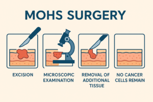
BCC develops most often in people with a long history of sun exposure or tanning bed use, fair skin, or a personal or family history of skin cancer. The good news: BCC grows slowly and almost never spreads to other parts of the body. Left untreated, though, it can still invade nearby tissue, nerves, or bone. Most BCCs are treated with a simple excision, but for BCCs on the face or other cosmetically sensitive areas, Mohs surgery for basal cell carcinoma is considered the gold standard, offering cure rates up to 99% while sparing the most healthy tissue. According to the American Cancer Society, basal and squamous cell cancers are highly curable when treated in their early stages.
Squamous Cell Carcinoma
Squamous cell carcinoma (SCC) is the second most common type of skin cancer, starting in the flat squamous cells found in the upper layers of the epidermis.
What Squamous Cell Carcinoma Looks Like
SCC often appears as a firm, red or skin-colored bump, a scaly patch, or a sore that doesn’t heal. Like BCC, it typically develops on sun-exposed skin (the face, ears, lips, neck, and backs of the hands), though it can also arise in old scars, chronic wounds, or areas of long-standing skin irritation.
Why It Needs Prompt Treatment
SCC is also generally slow-growing, but it carries a somewhat higher risk of spreading than BCC, particularly when it develops on the lips, ears, or scalp, or in patients with a weakened immune system, for example, transplant recipients who take anti-rejection drugs that suppress the immune system. Left untreated, SCC can grow more quickly and, in a small percentage of cases, spread to lymph nodes or beyond. Because of this, prompt evaluation of any new or non-healing sore is important, and treatment—often excision or Mohs surgery with local anesthetic—is generally straightforward when caught early.
Melanoma
Melanoma develops from melanocytes, the pigment-producing cells responsible for skin color. It’s the least common of the three major skin cancers, but it’s the one dermatologists watch most closely because melanoma can spread early.
The ABCDE Warning Signs
Melanoma often develops as a new dark spot and not from an existing mole. Dermatologists use the ABCDE rule to help patients spot early warning signs:
- Asymmetry (one half doesn’t match the other)
- Border irregularity (edges are notched or blurred)
- Color that varies within the same spot
- Diameter larger than a pencil eraser
- Evolution—any change in size, shape, or color over time
If you notice any of these, it’s worth having the spot evaluated; our guide to the warning signs of skin cancer covers self-exams in more detail.
Ugly Duckling Warning Sign
Melanoma often looks different than other normal moles on a person’s skin. This appearance is called “the ugly duckling sign,” meaning when compared to a person’s other moles, a melanoma looks different and stands out from the background moles. If you notice an “ugly duckling” mole, it’s important to get it checked out.
Why Melanoma Spreads Faster Than Other Types
Unlike BCC and SCC, melanoma can grow both across the surface of the skin and downward into deeper layers relatively quickly, giving it access to blood vessels and lymph nodes. That’s what allows it to metastasize to other organs if it isn’t caught early. According to the Skin Cancer Foundation, the five-year survival rate for melanoma detected early, before it spreads, is about 99%—which is exactly why early detection matters so much for this particular type.
What Is the Most Dangerous Type of Skin Cancer?
Of the common types of skin cancer, melanoma is considered the most dangerous—not because it’s the most common, but because of how quickly it can spread if left untreated. Basal cell and squamous cell carcinomas are far more common but rarely life-threatening when treated promptly.
| Skin Cancer Type | Relative Frequency | Metastasis Risk | 5-Yr Survival (Early Stage) |
| Basal Cell Carcinoma (BCC) | Most common (~80%) | Very low | >99% |
| Squamous Cell Carcinoma (SCC) | Second most common | Low, higher than BCC | ~95% |
| Melanoma | Less common | Higher if untreated | ~99% (drops if spread) |
| Merkel Cell Carcinoma | Rare | High | ~75% |
The takeaway isn’t to fear any particular type. It’s that any new, changing, or non-healing spot deserves a professional look, since the type of skin cancer you can’t identify yourself is the one that can do the most damage.
Less Common but Serious: Merkel Cell Carcinoma and Precancers
While basal cell carcinoma, squamous cell carcinoma, and melanoma make up the vast majority of diagnoses, they aren’t the only types worth knowing about.
Merkel cell carcinoma is rare (roughly 40 times less common than melanoma) but notably aggressive. It tends to appear as a fast-growing, firm, skin-colored or reddish-purple bump, most often in older adults or those with weakened immune systems. Despite its rarity, it’s the second-leading cause of skin cancer death after melanoma, according to the Skin Cancer Foundation, because it tends to spread quickly once it develops.
Actinic keratosis isn’t a skin cancer itself but a precancer—a rough, scaly, pink or red patch caused by sun damage that can develop into squamous cell carcinoma if left untreated. Because it’s so common and so treatable at this early stage, catching and treating actinic keratosis is one of the simplest ways to prevent a future skin cancer diagnosis altogether.
How Darabi Dermatology Diagnoses and Treats Skin Cancer
Getting an accurate diagnosis quickly matters, and that’s an area where our practice works differently than most. Dr. Darabi is one of very few dermatologists in the country who is triple board-certified in Dermatology, Mohs Micrographic Surgery, and Dermatopathology. That means when a spot is biopsied, it’s usually read by Dr. Darabi himself in our in-house dermatopathology lab—not sent to an outside lab you’ll never speak with—so in most cases there’s no gap between the person examining your skin and the person diagnosing it under the microscope.
Once a diagnosis is made, treatment is tailored to the type, size, and location of the skin cancer: simple excision for many low-risk basal and squamous cell carcinomas, Mohs surgery for cancers in cosmetically sensitive areas or with a higher risk of recurrence such as the face, ears, scalp, neck and hands, and coordinated care with oncology specialists for melanoma or Merkel cell carcinoma that requires it. Patients typically get in for an evaluation within one to two weeks, rather than the two-month wait common at many practices.
Frequently Asked Questions About Different Types of Skin Cancer
What are the 3 main types of skin cancer?
The three main types are basal cell carcinoma, squamous cell carcinoma, and melanoma. Basal cell and squamous cell carcinoma are the most common and rarely spread; melanoma is less common but could be more dangerous.
Which type of skin cancer is most common?
Basal cell carcinoma is the most common, accounting for about 80% of all skin cancer diagnoses.
Is melanoma really the most dangerous type of skin cancer?
Yes. While it’s far less common than basal cell or squamous cell carcinoma, melanoma is more likely to spread to other organs if not caught early, which makes it the most dangerous of the common types.
Can basal cell or squamous cell carcinoma spread to other organs?
It’s uncommon but possible, especially with squamous cell carcinoma in high-risk locations or in patients with a weakened immune system. Basal cell carcinoma very rarely spreads but can still cause significant local damage if untreated.
What does skin cancer usually look like?
It varies by type, but common signs include a pearly or waxy bump, a sore that doesn’t heal or keeps reopening, a scaly red patch, or a brown mole that’s changing in size, shape, or color.
How is skin cancer diagnosed?
A dermatologist examines the spot and, if it looks suspicious, performs a biopsy—removing a small sample of tissue to be examined under a microscope by a dermatopathologist.
Are there skin cancers besides the “big three”?
Yes. Merkel cell carcinoma is a rare but aggressive skin cancer, and actinic keratosis is a common precancer that can develop into squamous cell carcinoma if untreated.
How often should I get a skin cancer screening?
Most people benefit from an annual skin exam, though those with a personal or family history of skin cancer, many moles, or significant sun exposure may need more frequent checks.