By Kamruz Darabi, MD, FAAD, FASMS Triple board-certified dermatologist, Mohs skin cancer surgeon, and dermatopathologist at Darabi Dermatology, serving Chaska and Hutchinson, Minnesota.
Hearing that you need a skin biopsy can bring up a lot of questions and, understandably, some anxiety. The good news is that a biopsy is a routine, well-understood procedure, and it’s the only way to know for certain whether a spot is benign or cancerous. Understanding what to expect can make the whole process feel much less intimidating.
Why a Skin Cancer Biopsy Is Recommended
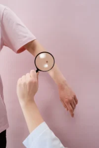
Your dermatologist may recommend a biopsy for skin cancer when a mole or skin lesion looks suspicious based on its appearance, size, color, or how it’s changed over time. This includes the classic ABCDE warning signs: asymmetry, irregular borders, uneven color, a diameter larger than a pencil eraser, or a spot that’s evolving, meaning it is changing in color, size or shape over time. A visual exam or dermoscopy exam alone can’t definitively confirm what’s going on. A biopsy removes a small sample of tissue so a pathologist can examine it under a microscope and determine whether it’s benign, precancerous, or one of the three main types of skin cancer: basal cell carcinoma, squamous cell carcinoma, or melanoma.
Types of Skin Cancer Biopsies
There isn’t just one way to do a skin biopsy. Your dermatologist chooses the method based on the suspected type of skin cancer, the lesion’s size and location, and how deep it may extend.
Shave biopsy:
The dermatologist shaves a thin layer from the top of the lesion. It’s quick, usually doesn’t require stitches, and is commonly used when a growth is thought to involve mainly the outer layer of skin.
Punch biopsy:
A small, round tool the diameter of a pea is used to remove a small circular “plug” of skin that goes deeper than a shave biopsy. This is used when a lesion may extend into the deeper layers of skin, and it’s often used when one or two sutures are planned as part of the biopsy itself.
Excisional and incisional biopsy:
A scalpel is used to remove the entire lesion (excisional) or a portion of a larger lesion (incisional), cutting through the full thickness of the skin. The wound is typically closed with two to three stitches. This method allows the deepest, most complete look at a lesion.
Melanoma Skin Biopsy: Why Technique Matters
When melanoma is suspected, the biopsy method carries extra weight. Melanoma staging and treatment planning depend heavily on how deep the tumor has grown into the skin, a measurement known as Breslow depth, measured in millimeters. Tumor depth is most accurately measured by evaluating the entire tumor via an excisional biopsy; other partial biopsy techniques, such as a partial shave or partial punch biopsy where only a portion of the lesion is sampled, are considered less accurate for this purpose. That’s why national guidelines list narrow excisional biopsy, such as a deep shave removal or small excisional biopsy, as the preferred technique for lesions suspicious for melanoma. The American Academy of Dermatology (AAD) and National Comprehensive Cancer Network (NCCN) currently list narrow excisional biopsy (fusiform excision) as the preferred technique for biopsying lesions suspicious for melanoma. It gives the pathologist the full picture needed to determine how far the melanoma has grown, what treatment is appropriate, and what the likelihood is of melanoma spreading beyond the original site.
What Happens During the Procedure
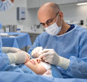
The procedure itself is straightforward and typically done right in the office. Your dermatologist will clean the area and inject a local anesthetic, which may cause brief stinging before the area goes numb. Once numb, you shouldn’t feel pain, though you may notice pressure or a tugging sensation as the sample is taken. Most biopsies take about 15 to 30 minutes from start to finish. Depending on the type of biopsy, the wound may be closed with a stitch or two, or left to heal with a simple bandage.
Aftercare and Healing
After your biopsy, some soreness at the site for a few days is normal, and over-the-counter pain relief with acetaminophen/Tylenol is usually enough to manage it. If stitches were placed, your dermatologist will let you know when they should come out, typically within one to two weeks. Keep the area clean with soap and water and follow any bandage or ointment instructions you’re given. Call your dermatologist if you notice bleeding that won’t stop with gentle pressure, or signs of infection like increasing redness, warmth, tenderness, or pus. A small scar at the biopsy site is expected regardless of which method was used.
What Happens With Results
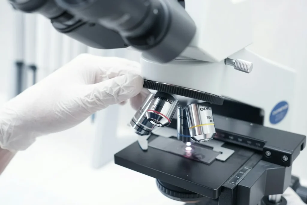
Once your sample is taken, it’s sent to a lab where a pathologist, often a dermatopathologist who specializes in skin tissue, examines it under a microscope. All Darabi Dermatology skin biopsies are examined by a board-certified dermatopathologist who specializes in examining only skin biopsies and nothing else. Turnaround time is typically about one to two weeks. If the results show skin cancer, your dermatologist will discuss what was found and walk you through next steps, which may range from a simple in-office removal to a more involved treatment plan depending on the type and stage.
Frequently Asked Questions
Does a skin cancer biopsy hurt?
You shouldn’t feel pain during the biopsy itself at all. A local anesthetic numbs the area first, though you may feel some pressure and touch during the sample collection.
How long does a skin biopsy procedure take?
Most skin biopsies take about 15 to 30 minutes and are done right in the dermatologist’s office.
What’s the difference between a shave biopsy and a punch biopsy?
A shave biopsy removes a thin layer from the top of the skin and usually doesn’t need stitches. A punch biopsy removes a deeper, circular sample and may require one or two stitches depending on its size.
Why is excisional biopsy preferred for melanoma?
Because melanoma staging depends on how deep the tumor has grown, and tumor depth is most accurately measured on an excisional biopsy specimen. Shave and punch biopsies that don’t remove the entire lesion can be less accurate for determining that depth.
How long does it take to get skin biopsy results?
Results typically take about one to two weeks, though timing can vary depending on the lab and the complexity of the case.
Will a skin biopsy leave a scar?
Yes, you should expect a small scar from any type of skin biopsy, though the size and visibility depend on the method used and the location on your body.
A skin cancer biopsy is one of the most important, and most routine, tools in diagnosing skin cancer early. Knowing what type of biopsy to expect, what the procedure involves, and how results are handled can take a lot of the uncertainty out of the process.
If you have a spot you’re concerned about, or you’ve had a biopsy and have questions about what comes next, the team at Darabi Dermatology is here to guide you through every step, from evaluation to diagnosis to treatment.